Home > Science > SEM
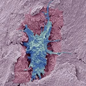
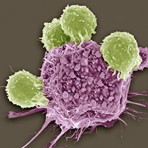
Coloured SEM of blood vessels in the skin

![]()
Wall Art and Photo Gifts from Science Photo Library
Coloured SEM of blood vessels in the skin
Skin blood vessels. Coloured scanning electron micrograph (SEM) of two small blood vessels (lower left and upper right) in the dermis of the skin. The dermis is the layer of the skin which contains blood vessels, sweat glands and hair follicles. The blood vessels are seen to contain red blood cells (red), which are responsible for carrying oxygen around the body to tissues which require it. Some white blood cells (round, green) are seen in the tissue surrounding the blood vessel at lower left. These cells help clear the body of infection. The area surrounding the blood vessels is largely connective tissue, which gives the skin its tone and elasticity. Magnification unknown
Science Photo Library features Science and Medical images including photos and illustrations
Media ID 6446497
© STEVE GSCHMEISSNER/SCIENCE PHOTO LIBRARY
Blood Blood Vessels Dermis Erythrocyte Red Blood Cell Skin Vessel Vessels White Blood Cell
FEATURES IN THESE COLLECTIONS
MADE IN THE USA
Safe Shipping with 30 Day Money Back Guarantee
FREE PERSONALISATION*
We are proud to offer a range of customisation features including Personalised Captions, Color Filters and Picture Zoom Tools
SECURE PAYMENTS
We happily accept a wide range of payment options so you can pay for the things you need in the way that is most convenient for you
* Options may vary by product and licensing agreement. Zoomed Pictures can be adjusted in the Cart.