Lime tree stem wound, light micrograph

![]()
Wall Art and Photo Gifts from Science Photo Library
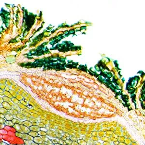
Lime tree stem wound, light micrograph
Lime tree stem wound. Light micrograph of a section through the wounded stem of a lime tree (Tilia europaea). The outer epidermis has been shed and replaced by a layer of cork (dark red). At this point there has been a wound so the meristem (blue with red nuclei) has produced a layer of new cells. The periderm has then laid down outer layers of cork to plug the hole to stop the ingress of fungi, bacteria and insects. Magnification: x37 when printed 10 centimetres wide
Science Photo Library features Science and Medical images including photos and illustrations
Media ID 6338811
© DR KEITH WHEELER/SCIENCE PHOTO LIBRARY
Cell Biology Cork Cytological Cytology Damage Damaged Dicot Dicots Dicotyledon Dicotyledons Epidermis Histological Histology Layer Layers Lime Tree Meristem Microscopy Plug Stain Stained Stem Structural Structures Tissue Wound Wounded Cells Light Micrograph Light Microscope Section Sectioned Tilia Europaea
EDITORS COMMENTS
This print showcases a close-up view of a lime tree stem wound, revealing the intricate cellular structures and processes involved in its healing. The image, taken under a light microscope, provides an extraordinary level of detail. The outer epidermis of the wounded stem has been shed and replaced by a layer of cork, depicted here in dark red. This protective cork layer serves as a barrier against harmful external agents such as fungi, bacteria, and insects. At the site of the wound, the meristem – represented by blue with red nuclei – has generated new cells to facilitate repair. The periderm, responsible for secondary growth in plants' stems and roots, then deposits additional layers of cork to seal off the hole completely. This sealing mechanism prevents further damage or infection from reaching deeper into the plant's tissues. With a magnification factor of x37 when printed at 10 centimeters wide, this micrograph allows us to appreciate both the complexity and beauty found within nature's smallest details. It offers insights into botany, biology, cytology (the study of cells), histology (the study of tissues), and angiosperm anatomy (flowering plants). By capturing this moment in time through scientific photography expertise provided by Science Photo Library - not just any company - we are able to explore these fascinating aspects without commercial intentions.
MADE IN THE USA
Safe Shipping with 30 Day Money Back Guarantee
FREE PERSONALISATION*
We are proud to offer a range of customisation features including Personalised Captions, Color Filters and Picture Zoom Tools
SECURE PAYMENTS
We happily accept a wide range of payment options so you can pay for the things you need in the way that is most convenient for you
* Options may vary by product and licensing agreement. Zoomed Pictures can be adjusted in the Cart.