Eustachian tube anatomy, artwork

![]()
Wall Art and Photo Gifts from Science Photo Library
Eustachian tube anatomy, artwork
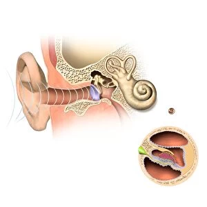
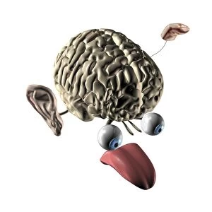
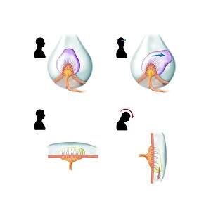
Eustachian tube anatomy. Artwork of the anatomy of the human Eustachian tube in its normal closed state (main artwork) and in an open state (inset, upper right). The Eustachian tubes (one for each ear) run from the middle ear to the throat. They are also called the auditory tube or pharyngotympanic tube. They allow drainage of fluids such as mucus, and pressure equalising. Methods of equalising pressure (called clearing the ears, white arrows) include yawning and swallowing. If pressure is not equalised, the result can be a burst ear drum. The ear drum is at centre in the main artwork, with the inner ear (ear bones, semi-circular canals, and cochlea) above and to its right
Science Photo Library features Science and Medical images including photos and illustrations
Media ID 9237997
© CLAUS LUNAU/SCIENCE PHOTO LIBRARY
Air Pressure Arrow Arrows Auditory Aural Closed Cochlea Cutaway Diagram Drainage Ear Canal Inner Ear Inset Internal Middle Ear Open Opened Otology Outer Ear Throat Tube Auditory System Cutouts Eustachian Tube Section Sectioned
EDITORS COMMENTS
This print showcases the intricate anatomy of the Eustachian tube, both in its normal closed state and in a visually striking open state. The main artwork highlights the central ear drum, surrounded by the inner ear's complex structures such as semi-circular canals and cochlea. Inset on the upper right corner, an additional image reveals this vital passage fully opened. The Eustachian tubes play a crucial role in our auditory system, connecting each middle ear to the throat. Also known as auditory or pharyngotympanic tubes, they serve multiple functions including fluid drainage and pressure equalization. To maintain equilibrium within these delicate organs, various methods are employed to clear ears and balance air pressure – illustrated by white arrows pointing towards yawning and swallowing. Failure to equalize pressure can have severe consequences like a burst eardrum. This comprehensive illustration emphasizes how essential it is for these tubes to function properly for optimal hearing health. With its clean white background and meticulous detailing, this artwork provides an insightful glimpse into one of our body's remarkable internal systems. It offers valuable information about otology (the study of ears) while simultaneously showcasing the beauty found within biological structures. Science Photo Library has expertly captured this anatomical masterpiece through their exceptional photography skills. This print serves as both educational material for medical professionals studying audiology or otolaryngology and a visually stunning piece that sparks curiosity about our own incredible biology
MADE IN THE USA
Safe Shipping with 30 Day Money Back Guarantee
FREE PERSONALISATION*
We are proud to offer a range of customisation features including Personalised Captions, Color Filters and Picture Zoom Tools
SECURE PAYMENTS
We happily accept a wide range of payment options so you can pay for the things you need in the way that is most convenient for you
* Options may vary by product and licensing agreement. Zoomed Pictures can be adjusted in the Cart.