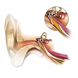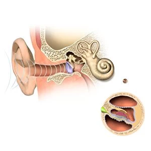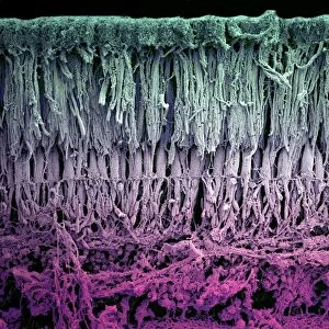
Human ear anatomy, artwork

![]()
Wall Art and Photo Gifts from Science Photo Library
Human ear anatomy, artwork
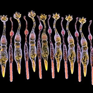
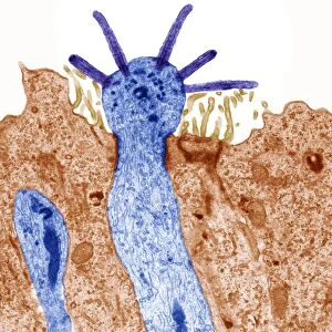
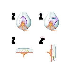
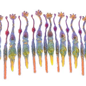
Human ear anatomy. Cutaway computer artwork showing the structure of the human ear, which consists of the outer ear (left), middle ear and inner ear (right). The ear drum (tympanic membrane, centre) separates the inner and middle ear, and transmits sound to the ossicles (centre right). This system of tiny bones transmits sound from the air-filled middle ear to the fluid filled cochlea (coiled, centre right) in the inner ear. The cochlea contains the organ of corti, which contains rows of hair cells topped with stereocilia. These cilia touch the tectorial membrane and detect tiny movements caused by sound-induced pressures in the inner ear fluids, which are translated into nerve impulses that travel to the brain, where they are deciphered as
Science Photo Library features Science and Medical images including photos and illustrations
Media ID 9207183
© BO VEISLAND/SCIENCE PHOTO LIBRARY
Audio Auditory Aural Auricle Bones Cochlea Detecting Detection Ear Drum Hearing Inner Ear Middle Ear Nerve Nerves Organ Of Corti Ossicle Ossicles Outer Ear Physiological Physiology Pinna Sense Sound Sounds Structures System Transmission Transmitting Tympanic Membrane Cells Cutouts
MADE IN THE USA
Safe Shipping with 30 Day Money Back Guarantee
FREE PERSONALISATION*
We are proud to offer a range of customisation features including Personalised Captions, Color Filters and Picture Zoom Tools
SECURE PAYMENTS
We happily accept a wide range of payment options so you can pay for the things you need in the way that is most convenient for you
* Options may vary by product and licensing agreement. Zoomed Pictures can be adjusted in the Cart.